Chest Pain
What to scan:
Findings
Use the corresponding finding images to identify what pathology your patient may have. Please remember that multiple diagnoses may exist at the same patient.
Normal:
Cardiac
Normal:
Cardiac


Pulmonary
Normal:
Pulmonary

IVC
Normal:
IVC


Acute MI:
Cardiac
Acute MI:
Cardiac


Diminished LVEF can suggest acute myocardial infarction
Pulmonary
Acute MI:
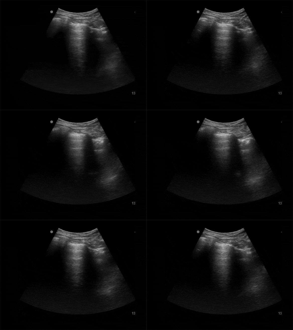
Pulmonary


Multifocal B-lines suggest congestive heart failure, in setting of acute MI this is high risk
IVC
Acute MI:
IVC
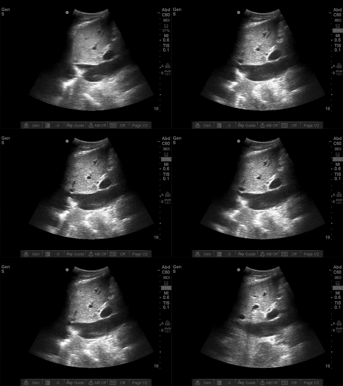

Fat IVC also suggests large MI
COPD/Asthma:
Cardiac
COPD/Asthma:
Cardiac
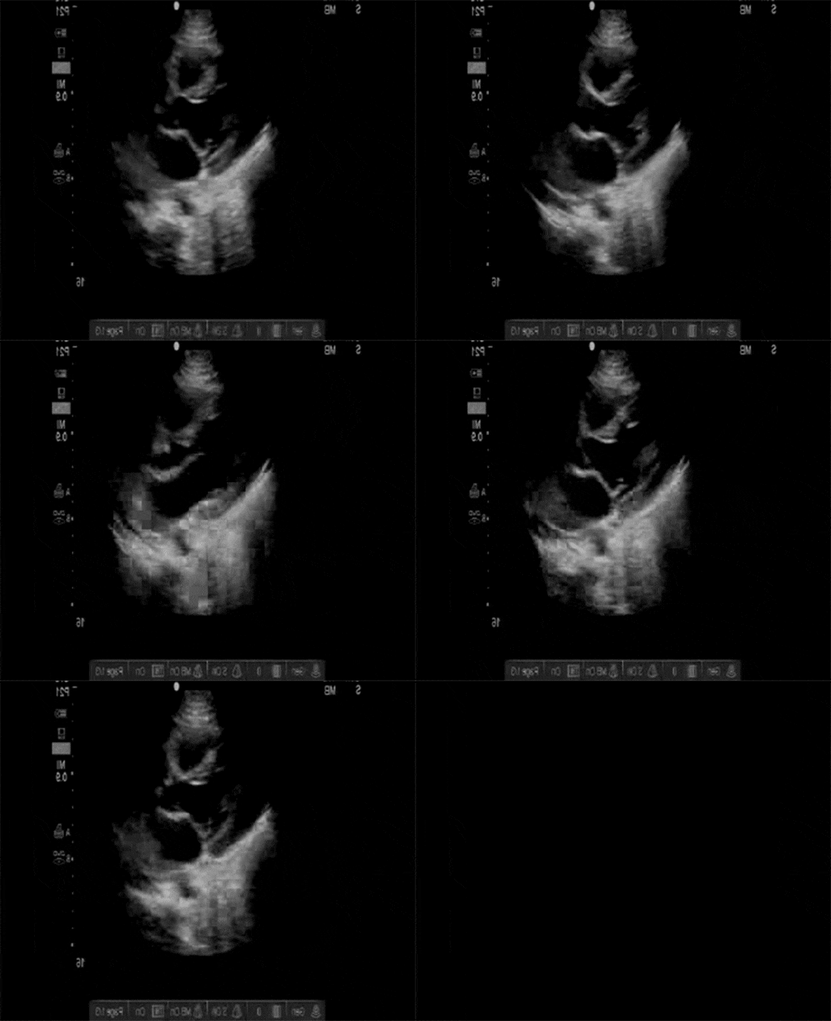

Normal to hyperdynamic LVEF
Pulmonary
COPD/Asthma:
Pulmonary


No B-lines
IVC
COPD/Asthma:
IVC
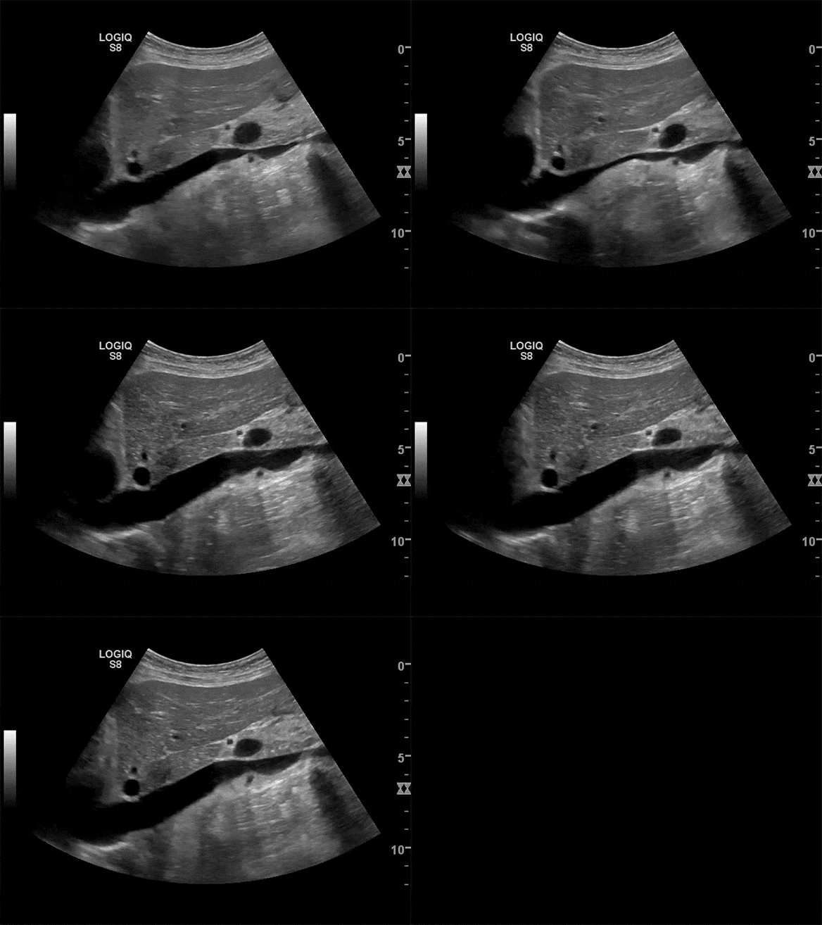

Normal to flat IVC
Pneumonia:
Cardiac
Pneumonia:
Cardiac


Normal to hyperdynamic LVEF
Pulmonary
Pneumonia:
Pulmonary


Focal B-lines to dense consolidation
IVC
Pneumonia:
IVC


Normal to flat IVC
Pericardial Effusion:
Cardiac
Pericardial Effusion:
Cardiac
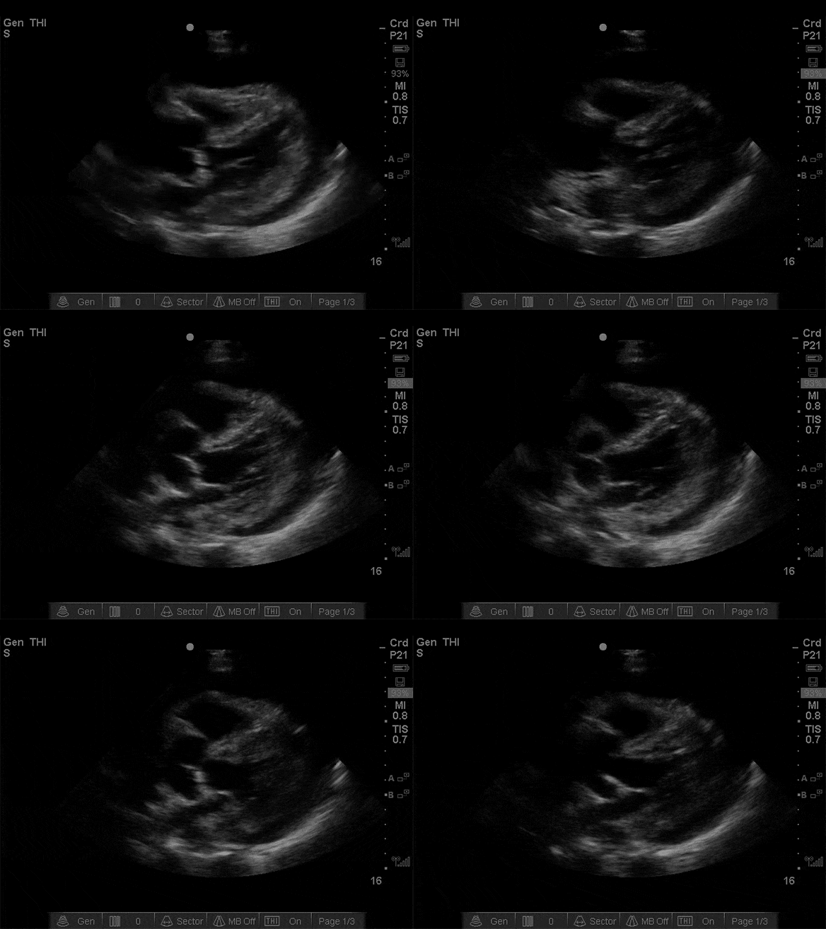

Normal to hyperdynamic LVEF
Pulmonary
Pericardial Effusion:
Pulmonary


Focal B-lines to dense consolidation
IVC
Pericardial Effusion:
IVC


Fat IVC
Pulmonary Embolism:
Cardiac
Pulmonary Embolism:
Cardiac


Large RV
Pulmonary
Pulmonary Embolism:
Pulmonary


No B-lines
IVC
Pulmonary Embolism:
IVC


Fat IVC
Pneumothorax:
Cardiac
Pneumothorax:
Cardiac


Variable LVEF
Pulmonary
Pneumothorax:
Pulmonary


No pleural sliding
IVC
Pneumothorax:
IVC


Fat IVC
Pleural Effusion
Cardiac
Pleural Effusion:
Cardiac


Variable LVEF
Pulmonary
Pleural Effusion:
Pulmonary


Large fluid collections above diaphragm
IVC
Pleural Effusion:
IVC


Variable IVC
Thoracic Aortic Dissection
Cardiac
Thoracic Aortic Dissection:
Cardiac


Wide aortic root, sometimes pericardial effusion
Pulmonary
Thoracic Aortic Dissection:
Pulmonary


Variable
IVC
Thoracic Aortic Dissection:
IVC


Variable
