Obtaining Cardiac Views
When to scan:
Where to put the probe:
4 Primary Cardiac Views:
• Parasternal Long Axis (PLAX)
• Parasternal Short Axis (PSAX)
• Apical 4 Chamber (A4CH)
• Sub Xiphoid
Improved views in left lateral decubitus with arm raised above head
Emergency/Radiology convention probe marker to patient right and indicator on screen left
Cardiology convention indicator will be on screen right
Use Parasternal Long Axis (PLAX) as primary view
Visualize PLAX as having 3 columns
Break view generation into 3 steps:
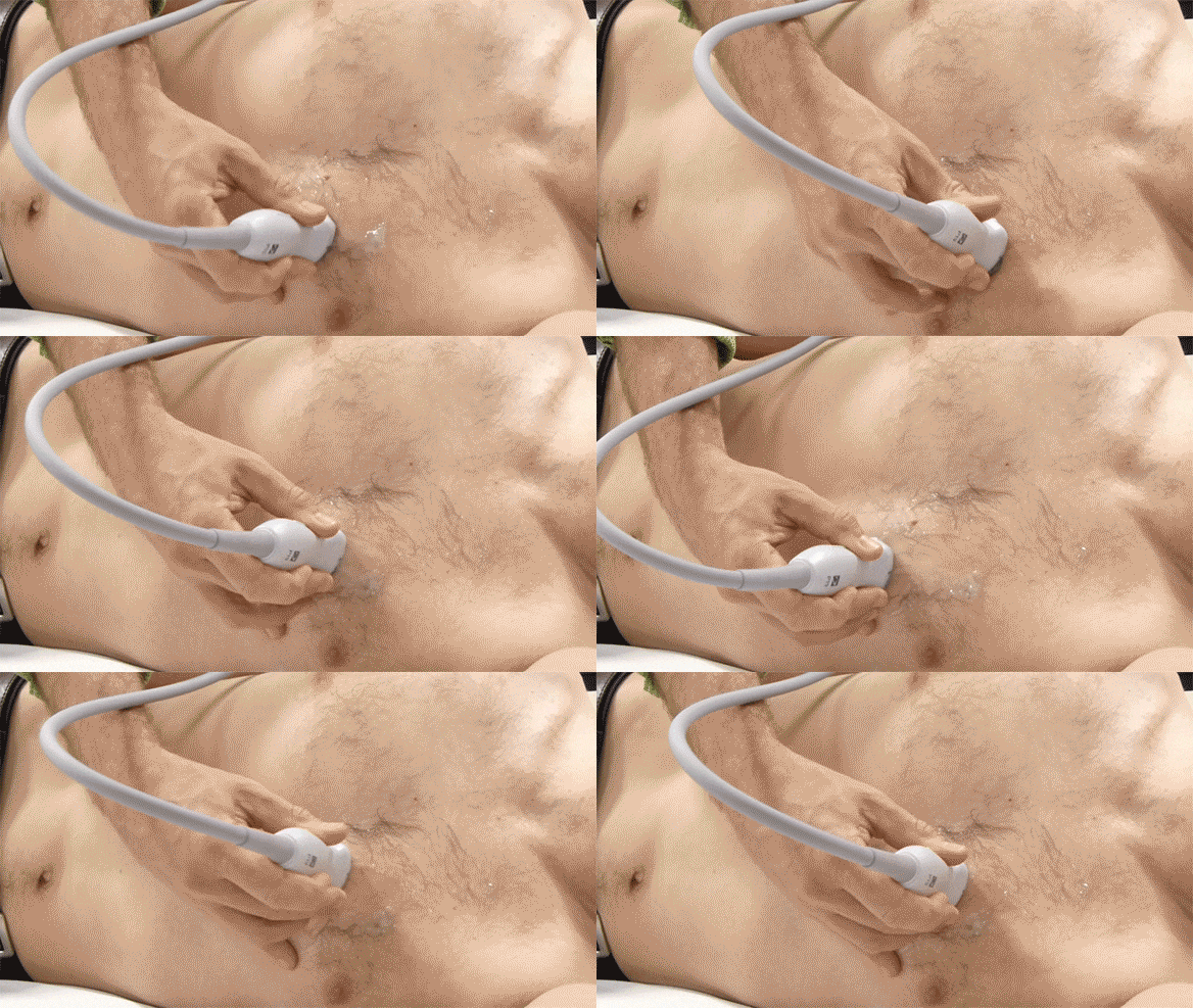
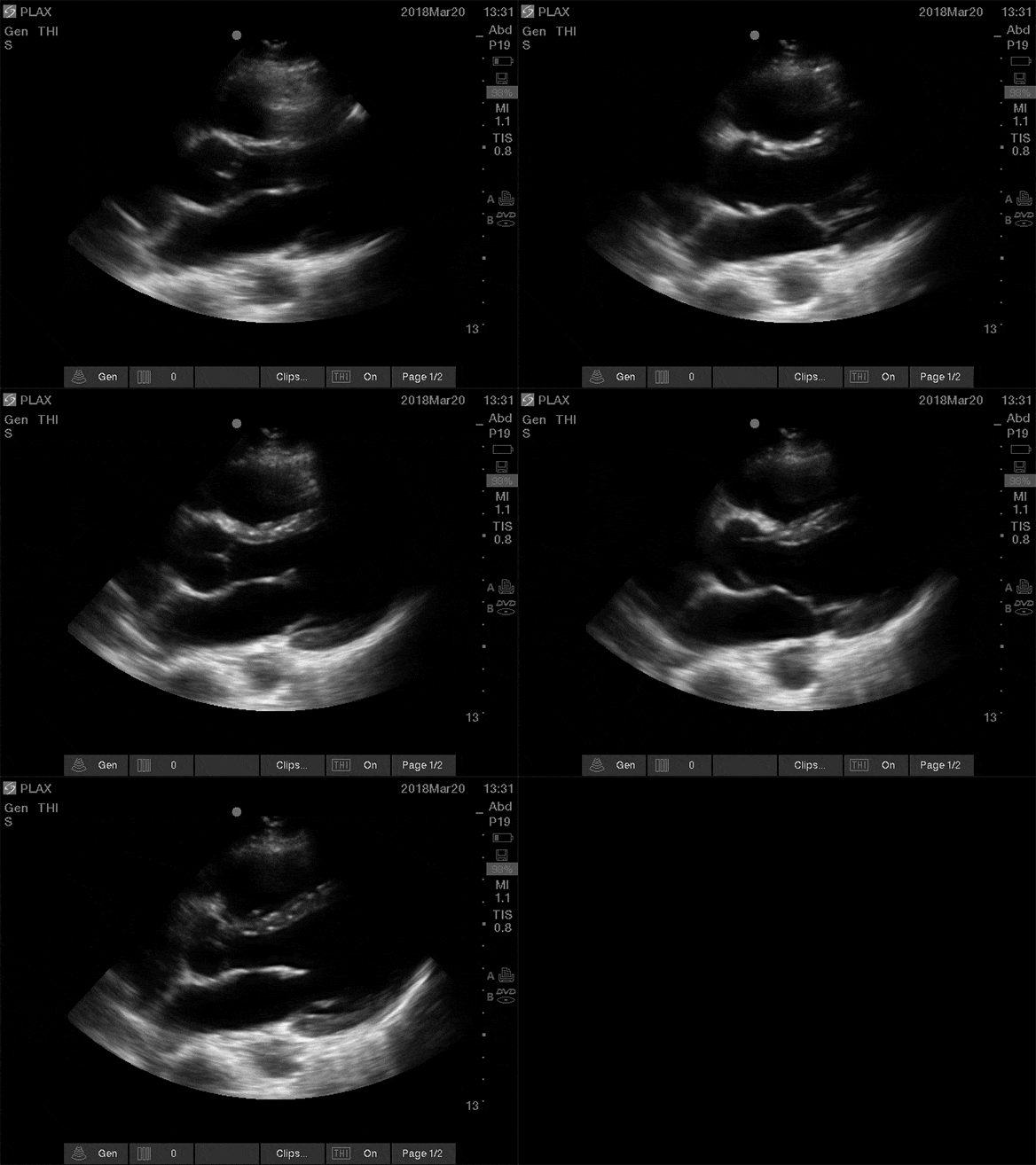
1. Window shopping:
• Indicator to right shoulder
• Move probe in arc to find best window
2. Fan for the middle (valves)
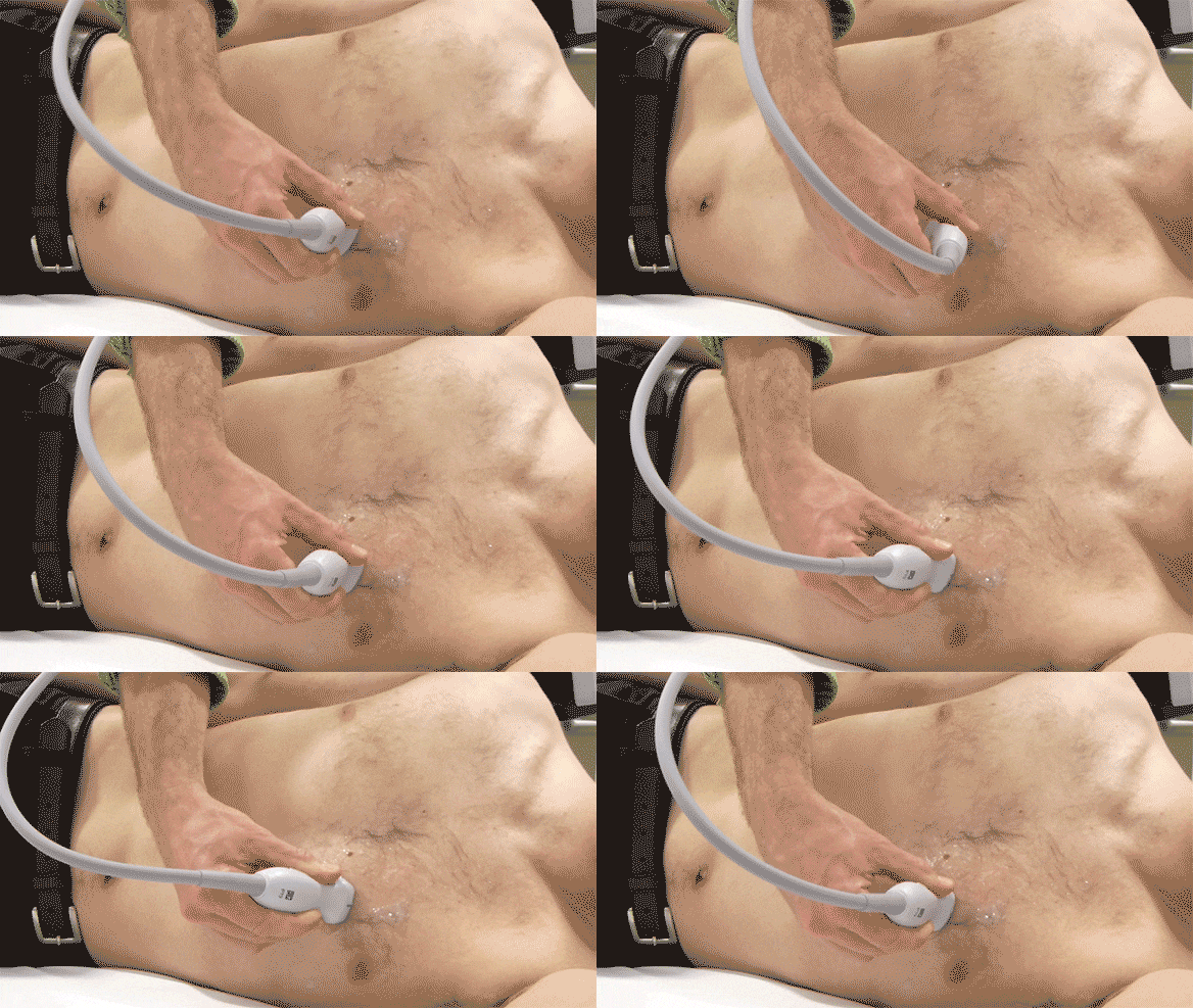
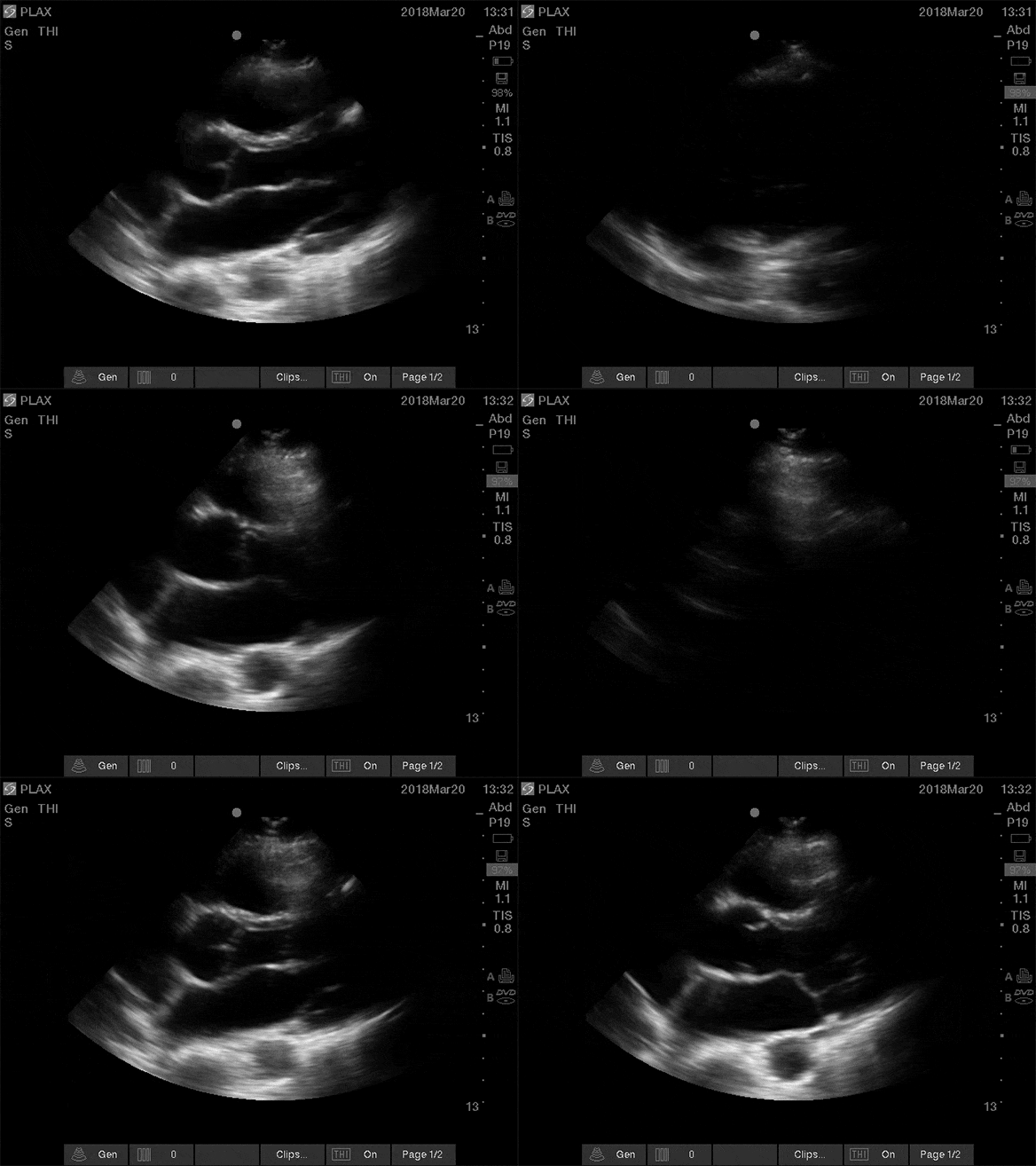
• Tilt/fan probe until valves are maximally opened
3. Rotate for the sides (LV)
• Keep angle from Step 2
• Rotate to maximally elongate the left ventricle


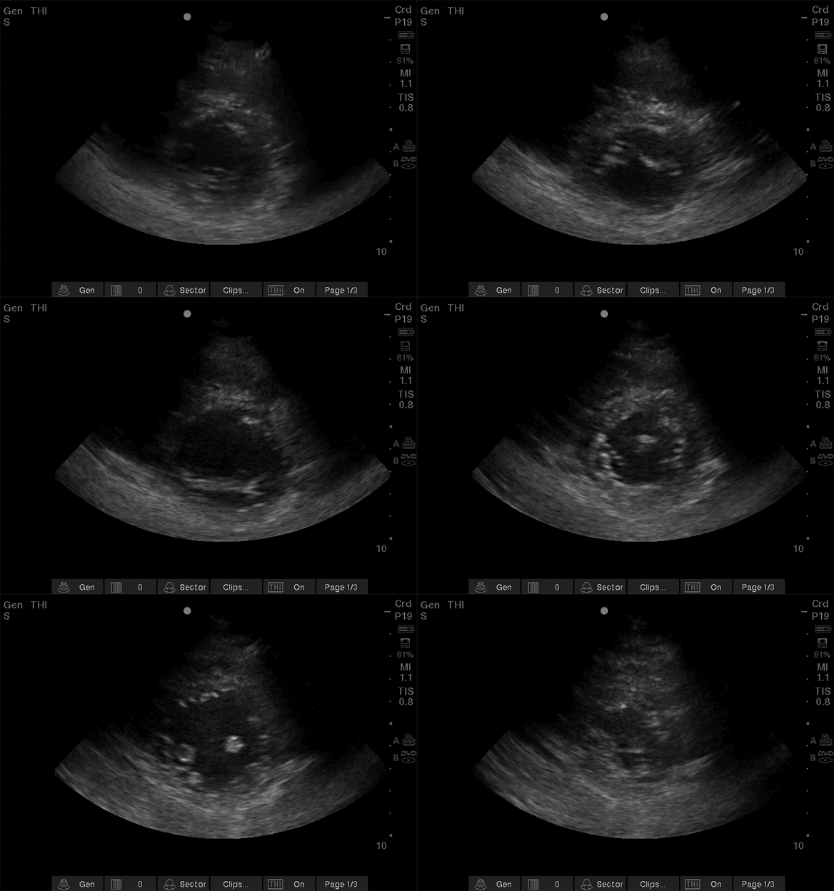
For Parasternal Short Axis:
1. From PLAX (mitral valve) rotate 90 degrees (counterclockwise)

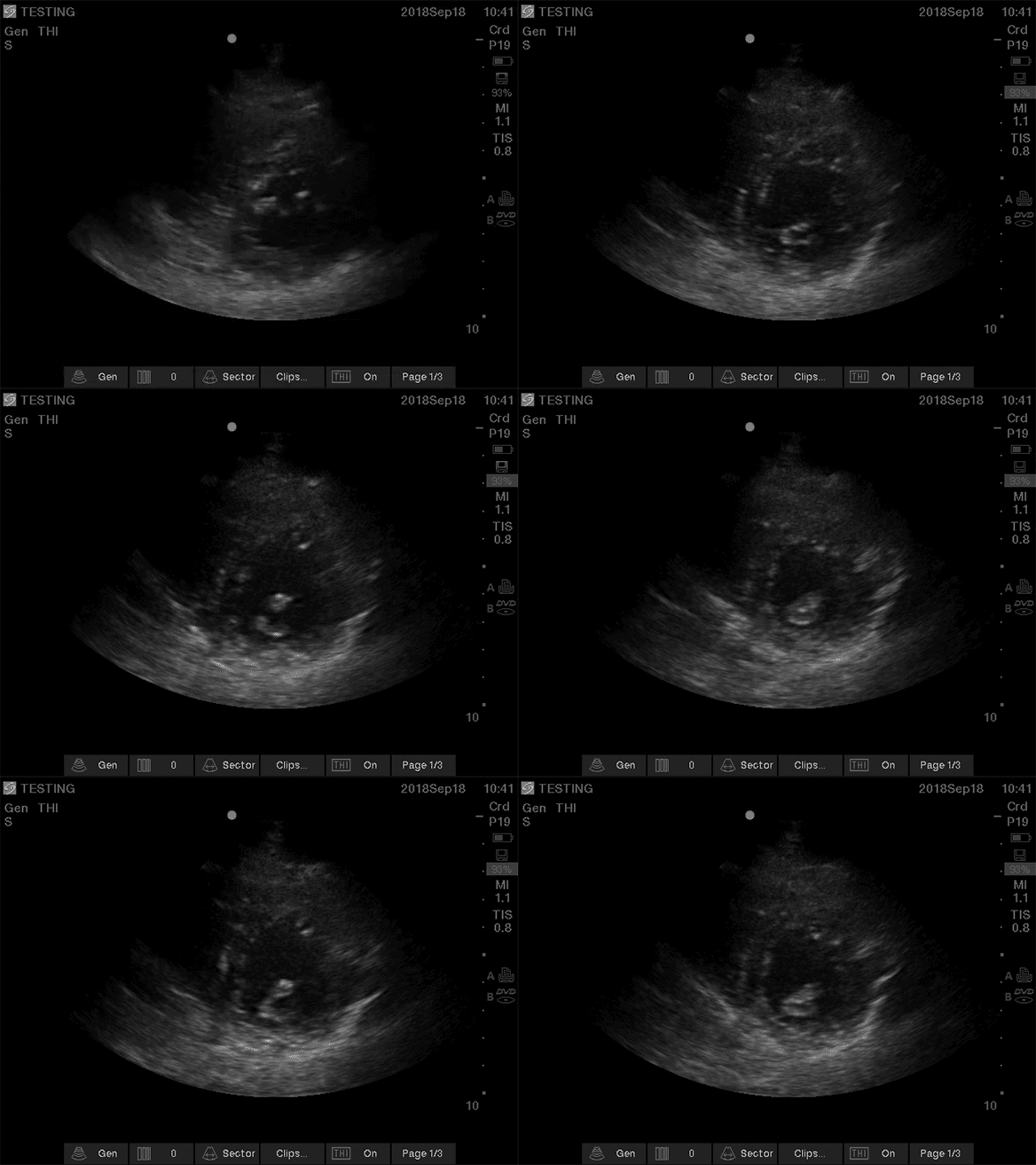
2. Slide towards apex to find papillary muscles
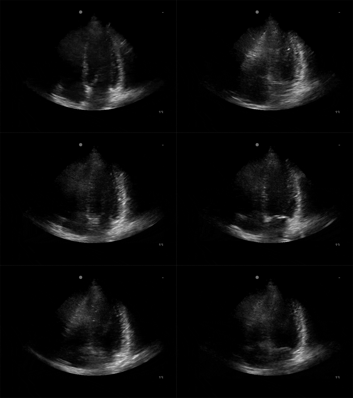
Apical 4 Chamber:
Probe marker to patient right. Start inferior and lateral to nipple line/inframammary fold. Window shop, fan, and rotate (as above to optimize image). Orient with vertical septum, x2 ventricles, and x2 atria in view.

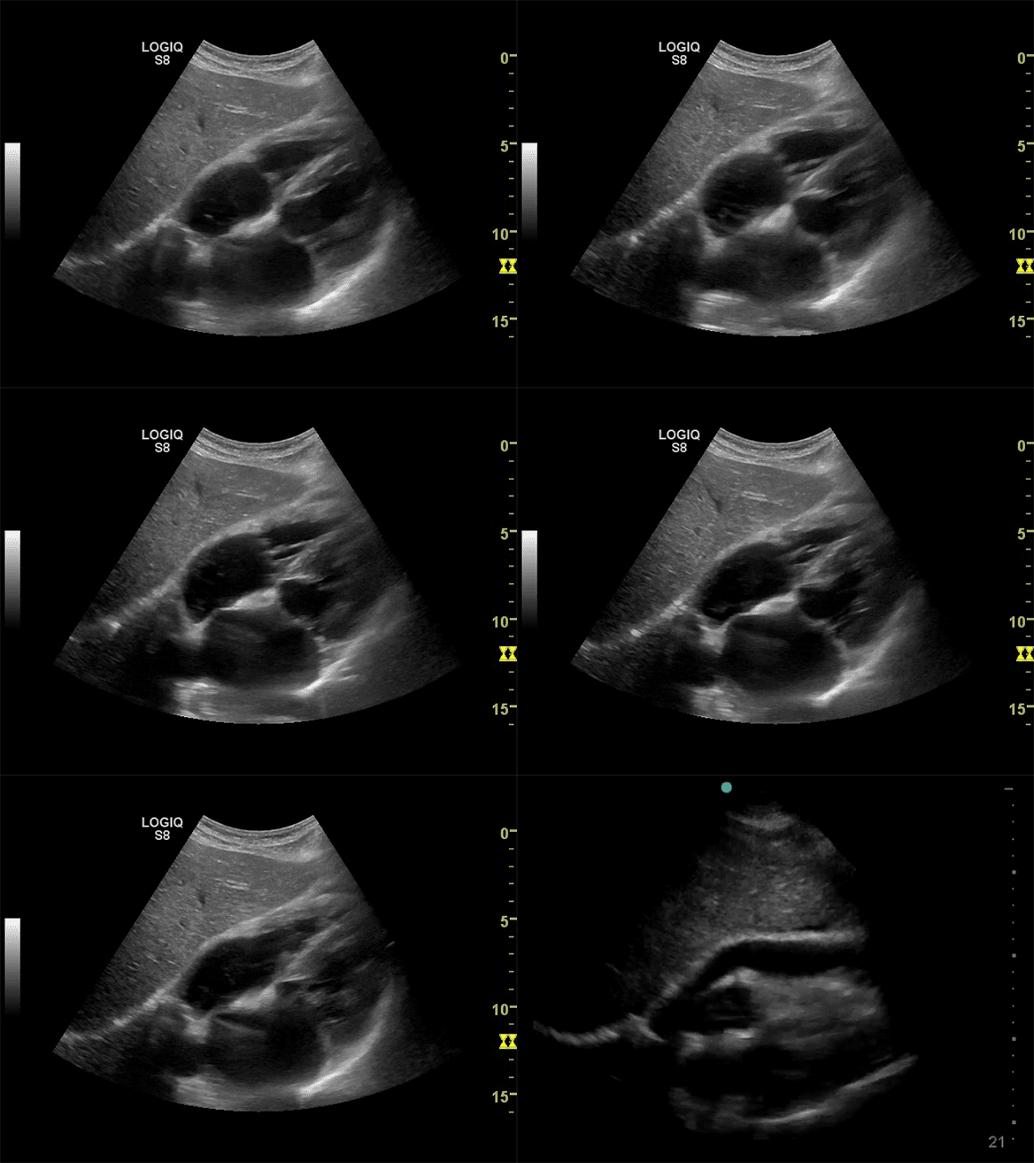
Sub Xiphoid View:
Start low in abdomen, parallel to floor and slowly move towards head, sweep through the heart.

4 Primary Cardiac Views:
• Parasternal Long Axis (PLAX)
• Parasternal Short Axis (PSAX)
• Apical 4 Chamber (A4CH)
• Sub Xiphoid
Improved views in left lateral decubitus with arm raised above head
Emergency/Radiology convention probe marker to patient right and indicator on screen left
Cardiology convention indicator will be on screen right
Use Parasternal Long Axis (PLAX) as primary view
Visualize PLAX as having 3 columns
Break view generation into 3 steps:
1. Window shopping:
• Indicator to right shoulder
• Move probe in arc to find best window


2. Fan for the middle (valves)
• Tilt/fan probe until valves are maximally opened


3. Rotate for the sides (LV)
• Keep angle from Step 2
• Rotate to maximally elongate the left ventricle


For Parasternal Short Axis:
1. From PLAX (mitral valve) rotate 90 degrees (counterclockwise)


2. Slide towards apex to find papillary muscles

Apical 4 Chamber:
Probe marker to patient right. Start inferior and lateral to nipple line/inframammary fold. Window shop, fan, and rotate (as above to optimize image). Orient with vertical septum, x2 ventricles, and x2 atria in view.


Sub Xiphoid View:
Start low in abdomen, parallel to floor and slowly move towards head, sweep through the heart.


